Accredited Imaging
Delivered by Skilled Professionals
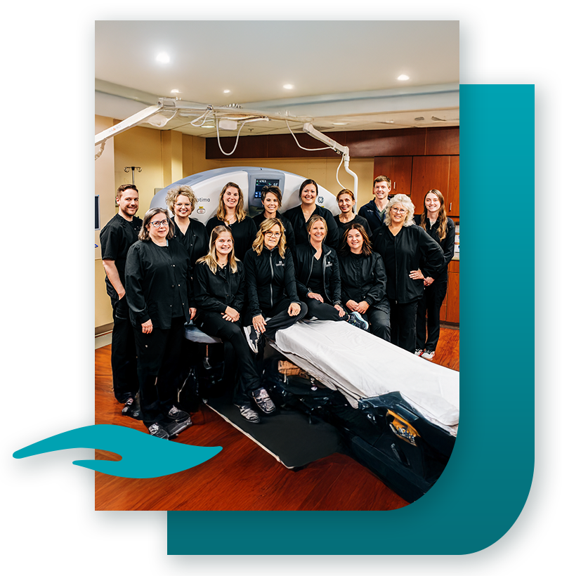
The Medical Imaging Department at Grand Lake Health System offers a full range of imaging services with cutting-edge technology and highly skilled healthcare professionals. Accredited by the American College of Radiology in
MRI, CT and Mammography.
Extended weekday hours are available.
Radiology & Imaging Services
3D/4D Ultrasound
Ultrasound (also called sonography or ultrasonography) is a painless imaging modality that uses high-frequency sound waves to create real-time images or video of the body’s internal structures. The images produced by ultrasound are viewed by a Radiologist (the doctor who reads the ultrasound) and can provide valuable information for diagnosing and directing treatment for a variety of diseases and conditions.
During an ultrasound a handheld device called a transducer or probe moves over an area of your body to obtain the images. A technologist applies a thin layer of gel to your skin so that the ultrasound waves can transmit from the transducer into your body obtaining images.
Bone Density (DEXA) Scan
DEXA is an exam that studies bone minerals to provide a bone density measurement and stands for Dual Energy X-ray Absorptiometry.
This test uses minimal radiation to determine the bone density of the spine, hip, or wrist. This test is highly sensitive and accurate and can diagnose bone loss associated with osteoporosis at an early stage. Procedure time: approximately 10 – 20 minutes
Inpatient and Outpatient Preparation Instructions:
Wear comfortable clothing such as a sweat suit – no metal zippers, buttons, or buckles.
DEXA exams are painless and only take 10 minutes to complete.
A patient history will be taken.
Family history of osteoporosis
X-ray findings of osteoporosis
Recent broken bones/fractures
Females post menopause or early menopause
Patients cannot have testing if:
- You have had a Barium study in the last 5 days.
- You are undergoing radioactive iodine thyroid therapy.
- You weigh over 270 lbs.
Risk Factors of Osteoporosis:
In the years following menopause, women will naturally experience bone loss because of a decrease in estrogen. Most will have no symptoms, even while the disease progresses. These factors add to your risk of developing osteoporosis.
- Age
- Smoking
- Caucasian or Asian Decent
- Inactive Lifestyle
- Certain Medications (including steroids & thyroid hormones)
- Thin or Small Build
- Previous Fracture
- Family History of Osteoporosis
- Alcohol Abuse
- Inadequate Calcium Intake
- Early Menopause
Why Physicians Request an Exam:
- To determine your actual bone density and fracture risk.
- To reveal early signs of bone loss with the fewest false positives or false negative results.
- To diagnose low bone mass that may signal the need for treatment.
Breast Imaging
The Women’s Imaging Center at Grand Lake Health System is committed to the health and well-being of every patient. Our experienced staff have been specially trained to guide patients efficiently and compassionately through every step of any process.
Services and procedures provided include:
- 3D Mammogram
- Stereotactic Breast Biopsy
- Breast Needle Localization
- Ultrasound Breast Cyst Aspiration
- Contrast-Enhanced Mammography
- MRI Breast
We understand that with any services, you want answers. At the Women’s Imaging Center, you’ll receive your diagnostic results the same day the tests are performed. Screening results will be provided as soon as they are available, and always within two weeks. When appropriate, you will meet with a radiologist who specializes in women’s imaging to address any concerns. We offer individual patient education, and we’ll assist you in accessing any additional resources you may require. For additional information, please call 419-394-3387, Ext. 3528.
CAT Scans
Computerized Axial Tomography (CAT) scan sometimes referred to as CT scan is a painless imaging modality that uses x-ray to create detailed images of the body’s internal structures. While lying on a flat table and using an x-ray tube that rotates around the body during scanning, cross sectional images or slices are produced. A CT scan can show detailed images of any part of the body including bones, soft tissue, organs, blood vessels and other internal structures. These images are and viewed by a Radiologists (the doctor who reads the x-rays) to help diagnose a patient’s condition.
CT scans may be done with or without Intravenous (IV) contrast and oral contrast. Contrast is used to help further identify internal structures and blood vessels. Your healthcare team will customize your CT scan based off your signs, symptoms and specific needs.
Fluoroscopy/General Radiography
Fluoroscopy is an imaging technique that uses x-rays to obtain real-time moving images of the body and is used for many types of exams. General Radiography is also called plain film radiography or X-Ray and is a used to create images in the internal structures in the body.
- Upper Gastro-Intestinal Series
- Barium Enema
- Voiding Cystogram
- I.V.P.
- Barium Swallow
- Modified Barium Swallow
- Hysterosalpingogram
- Routine Diagnostic X-rays
- Intravenous Pyelogram (I.V.P)
Interventional Radiology
Interventional Radiology is a medical specialty that performs various minimally-invasive procedures using different imaging guidance techniques such as fluoroscopy, CAT Scan, Ultrasound.
- Upper Gastro-Intestinal Series
- Barium Enema (3 Day)
- Barium Enema (1 Day)
- Voiding Cystogram
- I.V.P.
- Barium Swallow
- Modified Barium Swallow
- DEXA
- Hysterosalpingogram
- Joint Injections
- Lumbar Puncture
- Biopsy
- Paracentesis
- Thoracentesis
- Myelogram
- Abscess Drain
MRI
Magnetic Resonance Imaging (MRI) is an exam that uses a strong magnetic field and radio waves to visualize the body’s organs. If you are claustrophobic, you may want to ask your physician for medication to help you relax.
If any item applies to you in the following list, please inform the scheduling department:
- Surgical staples
- Metal implants
- Ear Prosthesis (implants)
- History of foreign body in eyes
- Aneurysm clips
- Dental Bridges
- Pacemaker
- Shrapnel (bullets)
- Hearing aids
- Pregnancy
- Inferior Vena Cava Filters
Patient Preparation Instructions:
- Follow your normal routine, eat normally, and take any medication you regularly take.
- Wear clothing with the least amount of metal as possible.
- No metal hairpins or barrettes.
- No hair products or make-up containing metallic flakes.
- Please bring your insurance cards.
- If your provider has ordered your exam With Contrast, you will be injected through an IV with a Contrast medium that is used to help highlight areas of interest on the MRI scan.
- The Radiologist (x-ray doctor) will “read” the scan and give the results to your doctor. Your doctor
- will share the results of this test with you.
For more information or questions, please call 419-394-3335, Ext. 3550.
Nuclear Medicine
Nuclear Medicine is a safe, specialized imaging technique that uses a small amount of radioactive material to help diagnose and monitor various conditions. Most scans involve a quick injection—typically through an IV or into the hand or arm—using a low-level radioactive substance that leaves the body within hours. The procedure is not painful, and side effects are rare. Scan times vary based on the test, with most lasting about an hour. Some scans may require specific preparation, and it’s important to inform your provider if you are pregnant or breastfeeding. Our team is here to ensure your comfort and safety every step of the way.
PET Scan
A positron emission tomography (PET) scan is an imaging test that helps reveal how your tissues and organs are functioning. A PET scan uses a radioactive drug (tracer) to show this activity. This scan can sometimes detect disease before it shows up on other imaging tests.
The tracer may be injected, swallowed or inhaled, depending on which organ or tissue is being studied. The tracer collects in areas of your body that have higher levels of chemical activity, which often correspond to areas of disease. On a PET scan, these areas show up as bright spots.
A PET scan is useful in revealing or evaluating several conditions, including many cancers, heart disease and brain disorders. Often, PET images are combined with CT or MRI scans to create special views. If you have any questions, please call 419-394-3335, Ext. 3550 for assistance.
More
Information
For more information regarding radiology and imaging services, please call 419-394-9509.
Certified Safe Sleep Champion
Grand Lake Health System achieved the highest certification level of Gold Safe Champion through our commitment to community leadership for best practices and education on infant safe sleep through the development of a hospital policy, staff training, parent education, implementing a wearable blanket program, becoming a Cribs for Kids partner, and a pledge to participate in ongoing audits and community outreach programs.
Click here to learn more about safe sleep.